3D Reconstruction from Biplanar X-Ray
This project is making high-quality 3D bone imaging accessible and affordable in resource-limited settings by developing AI systems that reconstruct 3D bone structures from orthogonal (biplanar) X-ray scans.
CT scans provide detailed 3D views of internal organs, crucial for diagnosis and trauma surgery, but are inaccessible or unaffordable in most rural and urban areas in countries like Nepal. Medical professionals rely on lower-cost, widely available X-ray scans, which do not provide a full 3D picture, leading to sub-optimal clinical outcomes and depriving many of quality care requiring 3D bone imaging.
Our goal is to make high-quality 3D imaging accessible and affordable using low-cost, readily available X-ray technology. We are developing an AI system that can take two (or very few) X-ray scans from different angles and accurately reconstruct 3D bone structures to improve diagnosis and surgical planning.
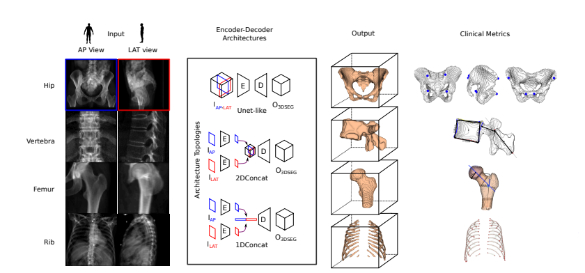
The project has benchmarked major AI methods using clinically relevant metrics, including bone morphometry parameters on major bones such as vertebra, pelvic bones, ribs, and femur. The platform includes reference implementations, APIs for data preprocessing, and automatic extraction of clinical landmarks and parameters. Early results show that attention-based models capturing global spatial relationships perform best, ribs are more challenging to reconstruct than other bones, and improvements in standard metrics (like dice score) do not always translate to clinical accuracy. This framework provides the foundation for selecting optimal models for real-world clinical deployment, guiding interventions such as fracture reconstruction and robust 3D imaging in low-resource environments.
NeurIPS Poster Presentation (2023)